We use cookies to make your experience better. To comply with the new e-Privacy directive, we need to ask for your consent to set the cookies. Learn more.
Hippo Pathway
Hippo-related products from Covalab
TAZ and YAP1
Yes-associated protein (YAP1) and Transcriptional coactivator with PDZ-binding motif (TAZ) are the main downstream mediators of the Hippo pathway.
YAP1/TAZ complex plays a crucial role in organ size control and tumour suppression by restricting proliferation and promoting apoptosis. Phosphorylation of YAP1/TAZ complex by LATS1 and 2 kinases blocks its translocation in to the nucleus and thus its transcriptional co-activator function on cellular genes, important for cell death, proliferation and migration.
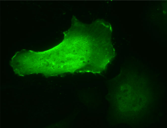
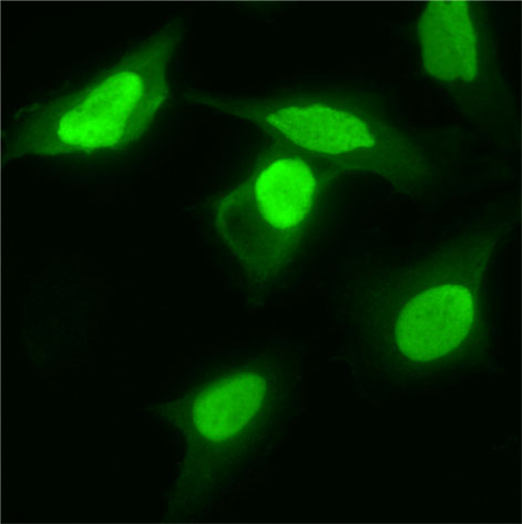
Anti-TAZ antibody IF staining of HeLa cells surexpressing TAZ protein.
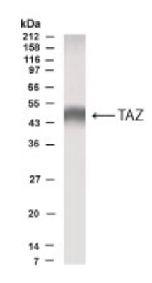
Anti-TAZ antibody WB staining of MDCK cells.
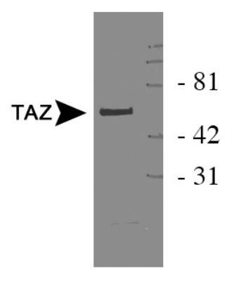
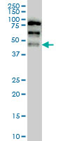
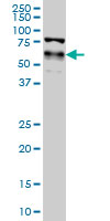
Anti-TAZ antibody WB staining of HEK 293 cell lysate.
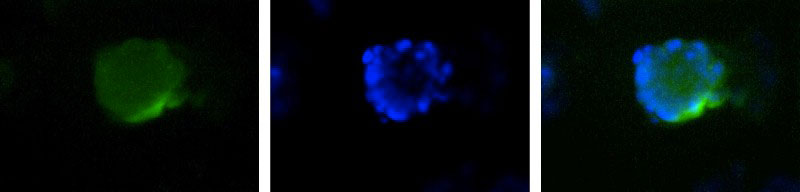
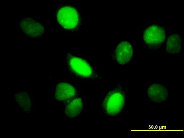
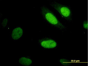
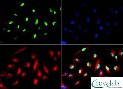
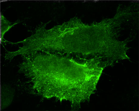
Anti-TAZ antibody ICC/IF staining of HepG2 cells (green). Nuclei (Blue) are counterstained with Hoecsht 33258.
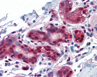
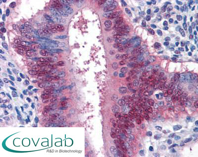
Anti-YAP1 antibody IHC staining of formalin-fixed, paraffin-embedded human breast.
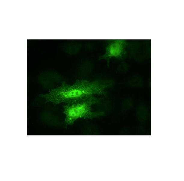
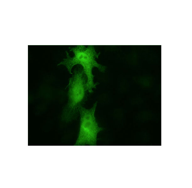
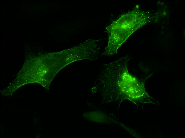
Anti-YAP1 antibody IF staining of HeLa cells.
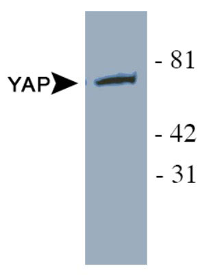
Anti-YAP1 antibody WB staining of HeLa cells lysates.
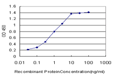
Detection limit for recombinant GST tagged YAP1 is approximately 0.03 ng/ml as a capture antibody.
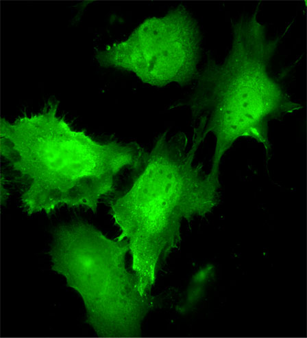
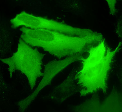
Anti-YAP1 antibody IF staining of HeLa cells.
Anti-YAP1 antibody IHC staining of formalin-fixed, paraffin-embedded human uterus.
Anti-YAP1 antibody WB staining of HeLa NE cell lysates.
Anti-YAP1 antibody WB staining of HEK 293 cell overexpressing YAP1.
Anti-YAP1 antibody IF staining of HeLa cells (green). Alpha tubulin and nuclei were counterstained with Dylight 550 (red) and DAPI (blue), respectively.
Other core proteins
The core of the Hippo pathway also includes a kinase cascade starting by MST1 and MST2, which phosphorylate and activate LATS1 and LATS2 kinases. LATS1 and 2 phosphorylate the YAP1/TAZ complex, which maintains it within the cytoplasm. Upon dephosphorylated conditions, this complex is translocated into the nucleus and binds DNA in complex with TEAD1 to TEAD4 (TEA domain-containing-sequence-specific transcription factors).
Anti-Serine/threonine-protein kinase LATS2 antibody IF stainings of HeLa cells transfected with Serine/threonine-protein kinase LATS2.
Anti-MST-2 antibody IF staining of HeLa cells transfected with MST-2.
Anti-TEAD-2 antibody IF staining of human breast tumors cells treated (+) or not (-) three days of TGFβ.

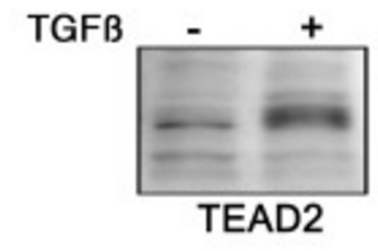
Anti-TEAD-2 antibody WB staining of human breast tumors cells treated (+) or not (-) three days of TGFβ.
Anti-TEAD-4 antibody IF staining of HeLa cells overexpressing TEAD-4.